
Publications

- Home
- Publications
- Research Publications
- Quantitative CRFF-OCT Imaging Features for Characterization of Disease Activity in Non-Segmental Vitiligo: A Machine Learning–Assisted Study
Quantitative CRFF-OCT Imaging Features for Characterization of Disease Activity in Non-Segmental Vitiligo: A Machine Learning–Assisted Study
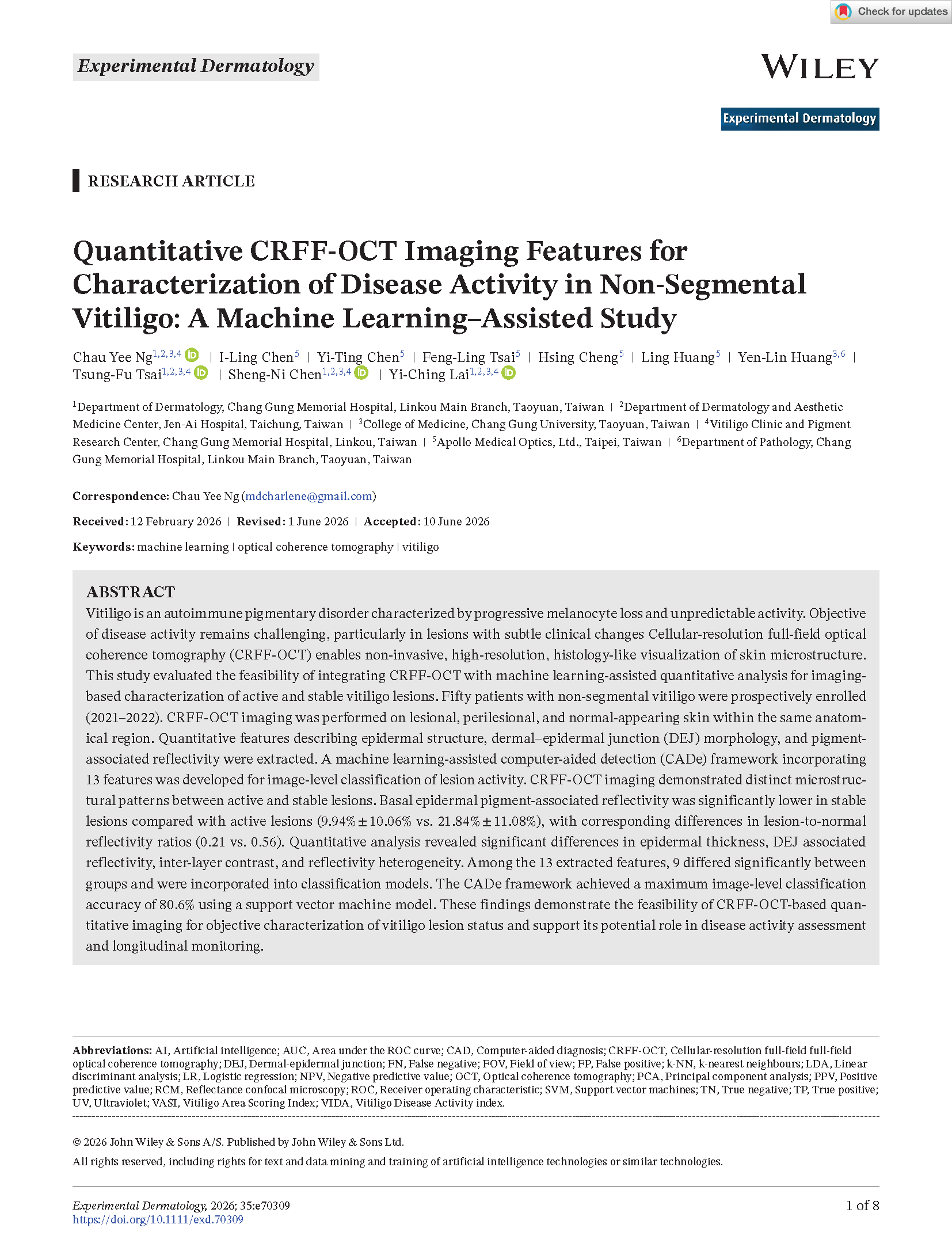
Vitiligo is an autoimmune pigmentary disorder characterized by progressive melanocyte loss and unpredictable activity. Objective
of disease activity remains challenging, particularly in lesions with subtle clinical changes Cellular-resolution full-field optical coherence tomography (CRFF-OCT)enables non-invasive,high-resolution,histology-like visualization of skin microstructure.
This study evaluated the feasibility of integrating CRFF-OCT with machine learning-assisted quantitative analysis for imaging-based characterization of active and stable vitiligo lesions. Fifty patients with non-segmental vitiligo were prospectively enrolled (2021–2022). CRFF-OCT imaging was performed on lesional, perilesional, and normal-appearing skin within the same anatomical region. Quantitative features describing epidermal structure, dermal–epidermal junction (DEJ) morphology, and pigment-associated reflectivity were extracted. A machine learning-assisted computer-aided detection (CADe) framework incorporating 13 features was developed for image-level classification of lesion activity. CRFF-OCT imaging demonstrated distinct microstructural patterns between active and stable lesions. Basal epidermal pigment-associated reflectivity was significantly lower in stable lesions compared with active lesions (9.94% ± 10.06% vs. 21.84% ± 11.08%), with corresponding differences in lesion-to-normal reflectivity ratios (0.21 vs. 0.56). Quantitative analysis revealed significant differences in epidermal thickness, DEJ associated reflectivity, inter-layer contrast, and reflectivity heterogeneity. Among the 13 extracted features, 9 differed significantly between groups and were incorporated into classification models. The CADe framework achieved a maximum image-level classification accuracy of 80.6% using a support vector machine model. These findings demonstrate the feasibility of CRFF-OCT based quantitative imaging for objective characterization of vitiligo lesion status and support its potential role in disease activity assessment and longitudinal monitoring.
of disease activity remains challenging, particularly in lesions with subtle clinical changes Cellular-resolution full-field optical coherence tomography (CRFF-OCT)enables non-invasive,high-resolution,histology-like visualization of skin microstructure.
This study evaluated the feasibility of integrating CRFF-OCT with machine learning-assisted quantitative analysis for imaging-based characterization of active and stable vitiligo lesions. Fifty patients with non-segmental vitiligo were prospectively enrolled (2021–2022). CRFF-OCT imaging was performed on lesional, perilesional, and normal-appearing skin within the same anatomical region. Quantitative features describing epidermal structure, dermal–epidermal junction (DEJ) morphology, and pigment-associated reflectivity were extracted. A machine learning-assisted computer-aided detection (CADe) framework incorporating 13 features was developed for image-level classification of lesion activity. CRFF-OCT imaging demonstrated distinct microstructural patterns between active and stable lesions. Basal epidermal pigment-associated reflectivity was significantly lower in stable lesions compared with active lesions (9.94% ± 10.06% vs. 21.84% ± 11.08%), with corresponding differences in lesion-to-normal reflectivity ratios (0.21 vs. 0.56). Quantitative analysis revealed significant differences in epidermal thickness, DEJ associated reflectivity, inter-layer contrast, and reflectivity heterogeneity. Among the 13 extracted features, 9 differed significantly between groups and were incorporated into classification models. The CADe framework achieved a maximum image-level classification accuracy of 80.6% using a support vector machine model. These findings demonstrate the feasibility of CRFF-OCT based quantitative imaging for objective characterization of vitiligo lesion status and support its potential role in disease activity assessment and longitudinal monitoring.
FAQ Classification
Related FAQ
- Recurrence and relapse in nonsegmental vitiligo: phenotypic and autoimmune predictors from a large retrospective cohort
- A comparative study of an advanced skin imaging system in diagnosing facial pigmentary and inflammatory conditions
- From zero to one: Recent advances in the pathogenesis, diagnosis, and treatment of vitiligo.
- Skin Interstitial Fluid and Plasma Multiplex Cytokine Analysis Reveals IFN-γ Signatures and Granzyme B as Useful Biomarker for Activity, Severity and Prognosis Assessment in Vitiligo
- Targeting the elevated IFN - γ in vitiligo patients by human anti - IFN - γ monoclonal antibody hampers direct cytotoxicity in melanocyte
- Pathogenic autoantibodies to IFN-γ act through the impedance of receptor assembly and Fc-mediated response
- Feasibility of Oral Tranexamic acid (TA) for Vitiligo Patients with Melasma
- A comparative study of suction blister epidermal grafting and automated blister epidermal micrograft in stable vitiligo
- Efficacy and safety of automated epidermal micrograft in patients with stable segmental and nonsegmental vitiligo
- Feasibility of High-Cellular-Resolution Full-Field, Artificial-Intelligence-Assisted, Real-Time Optical Coherence Tomography in the Evaluation of Vitiligo: A Prospective Longitudinal Follow-Up Study.